

The mission of QBRI's Microscopy and Imaging Core is to offer researchers efficient, reliable and innovative imaging solutions with the highest standards of instrumentation, accuracy, quality control and professional expertise.
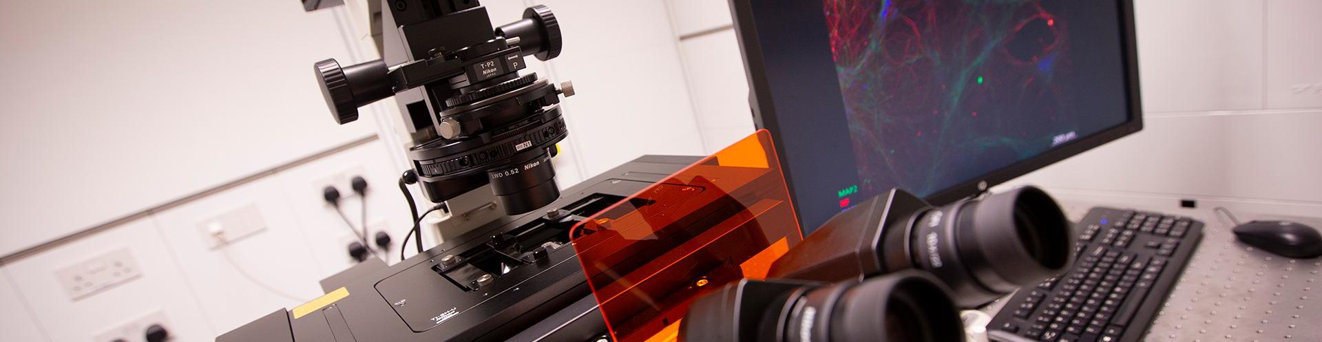
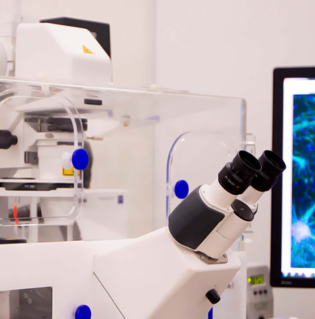
This is achieved by providing access to state-of-the-art instrumentation and the expertise of highly skilled professionals in the fields of microscopy and imaging. The unit is equipped with the most advanced and automated digital microscopy and live-cell imaging instrumentation.
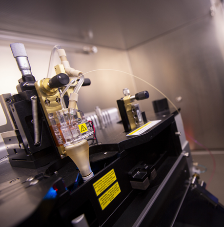
These technologies allow investigators to perform a wide variety of imaging research experiments that help them answer important biomedical questions related to their field of study. The core also provides training and easy access to researchers as well as services that range from routine microscopy to cutting-edge live-animal multi-photon microscopy.
The core’s expertise and services cover advice and support in the planning and evaluation of histopathology experiments, tissue processing and sectioning, cryopreservation, histological staining methods and laser caption microdissection microscopy.
Resources and Services
- Nikon A1+ MultiPhoton microscope
- Zeiss LSM 780 confocal microscope inclusive of incubation chamber
- Leica GSD super resolution microscope
- Zeiss Primo Star upright microscope
- Zeiss Axio Imager Z2 upright + camera fully automated fluorescence microscope
- Olympus Inverted microscope IX73 and IX83
- Zeiss Axio lmager A2 Microscope
- Several Olympus microscopes and Phase Contrast microscopes (Upright and Inverted)
Services – Optical Microscopy and Digital Imaging
- State-of-the-art microscopes for a wide variety of imaging research experiments
- Cutting-edge bright-field, dark-field and fluorescence imaging
- Standard, confocal and multi-photon microscopy applications
- Automated digital microscopy
- Advanced high-end live-cell imaging
- Image processing and analysis tools
- Training and education in microscopy and imaging
- Enhanced imaging research
- Support with data analysis and presentation
- Data management and storage
- Digital whole slide scanner, Leica Aperio® AT2 Turbo
- High quality digital slide scanner, Leica Aperio CS2
- Automated upright microscope, Leica DM4000B
- Semi-motorized rotary microtome, Leica RM2245
- Cryostat, Leica Biosystems CM3050 S
- Bright Instrument 8000 Sledge (Sliding Microtome
- Leica rotary microtome RM2125
- Leica ASP6025 Automated Tissue Processor
- Leica EG1150 modular tissue embedding center
- Leica Biosystems ST5020 Multistainer
- CryoViz robotic sectioning and imaging system
- Dako Omnis automated staining System GI100
- Dako Coverslipper
Services – Histopathology
- Tissue processing, embedding, and sectioning
- Cryopreservation and cryosectioning (Leica Cryostat and Microtome systems)
- Different histological staining methods, such as H&E and special stains
- Immunohistochemical and immunofluorescence techniques
- Laser Capture Microdissection microscopy (Leica)
- Expert advice in the planning and evaluation of histopathology experiments
Flow Cytometry
The mission of the QBRI Flow Cytometry Core is to offer researchers efficient, reliable and innovative flow cytometry and cell sorting solutions with the highest standards of instrumentation, quality control, biosafety and productivity. This is achieved by providing access to state-of-the-art instruments and professional flow cytometry and cell sorting services and is supported by highly motivated staff with extensive skills and profound expertise in the field of flow cytometry.
In addition, the flow cytometry core provides support and guidelines with regards to flow cytometry education, operation of instruments, software workflows and Biosafety to fulfill the mission of a fruitful, collaborative, and highly innovative environment for research and development.
Resources and Services
- BD Accuri C6 flow cytometer (2 lasers, 6 detectors)
- BC Gallios flow cytometer (3 lasers, 12 detectors)
- BD LSR Fortessa X-20 SORP flow cytometer (5 lasers, 20 detectors)
- Amnis Image Stream MKII imaging flow cytometer (2 lasers, 6 detectors)
- CellSee CTC Single Cell Analysis System
- 2 x BD FACSAria SORP cell sorter (5 lasers, 18 detectors)
- BD FACSJazz cell sorter (3 lasers, 8 detectors, in BSL-2 cabinet)
- Flow cytometry software analysis (BD FACSDiva, FlowJo, BD Sortware, ModFit LT, etc.)
- Advice on sample preparation (reagent selection, protocol design & optimization)
- Support multicolor panel design (negative controls, compensation setup, and multicolor panels)
- Multicolor sample acquisition and analysis
- Single cell analysis and sorting applications
- Aseptic cell sorting and biosafety advice
- Surface phenotyping and intracellular staining
- Apoptosis, cell cycle analysis, and proliferation
- Flow cytometry training and education of researchers and students
- Support with data analysis and presentation
- Data management and storage